
Medically reviewed by: Health is Heaven Medical Review Board | Published by Ganesh G Kamble, Health is Heaven | Published: April 22, 2026 · Last updated: October 16, 2026
The wellness industry frequently treats intermittent fasting as a generic weight-loss switch, casually recommending that eating dinner slightly earlier guarantees biological optimization. This fundamentally misunderstands human cellular architecture. If you are just starting out, reading our intermittent fasting for beginners guide can establish a solid physiological foundation before pursuing deeper fasts. The overarching goal of fasting is not simply burning stored abdominal fat; the target is profound internal recycling. Millions of individuals look for the exact fasting hours for autophagy timeline details, only to be fed highly inaccurate, diluted timelines. You cannot trigger a massive intracellular cleanup by simply skipping breakfast. Autophagy is a severe, survival-based stress response; it must be forcefully mandated through precise chronological engineering.
Autophagy typically begins around 16-24 hours into a fast and deepens progressively from 24 to 48+ hours, though the exact onset varies with factors like prior diet, glycogen stores, and activity level. The hour-by-hour breakdown below explains what is happening in your cells at each stage.
At healthisheaven.com, we approach the human body exactly as we map a highly complex data server network. The mTOR pathway (Mechanistic Target of Rapamycin) is your body's primary growth and building algorithm. As long as any amino acids or glucose are actively circulating in your upper digestive tract, mTOR is permanently switched on, continuously pushing cells to grow. If a cell becomes corrupted, oxidized, or prematurely aged (senescent), mTOR blindly continues to feed it. To stop this toxic growth and force the system to hunt down and cannibalize these weak, malfunctioning cells for spare parts (Autophagy), you must definitively cut the power supply. You must mathematically starve the mTOR pathway to force it entirely offline. To achieve cellular repair, you must understand the exact fasting hours deep autophagy requires to activate, transition, and complete this cellular purge.
To initiate an effective fasting and cellular rejuvenation plan, it is vital to establish your baseline metabolic metrics. Utilizing our free tdee calculator allows you to evaluate your current energy expenditure and track body hydration changes accurately during the fast. Before we dissect the biological timeline in detail, you can run your numbers in our clinical tools below to establish your hydration parameters and baseline caloric needs.
Calculate Hydration and Metabolic Metrics for Safe Fasting
Fasting triggers a massive release of cellular waste that must be filtered by the kidneys. Use our clinical calculators to estimate your mandatory fluid requirements, evaluate baseline energy levels, and check your biological age targets before executing a deep fast.
1. How Long Do You Need to Fast for Autophagy? (Hour-by-Hour Timeline)
To utilize fasting effectively for cellular rejuvenation, you must understand the exact biological thresholds your body crosses over a continuous timeline. Autophagy is not a binary switch; it is a progressive physiological response that increases as hepatic energy reserves are depleted. The timeline below outlines the metabolic and cellular transitions that occur during a prolonged fast, based on clinical endocrinology and kinetic studies conducted through 2026.
Hours 0 to 12: The Glycogen Depletion Phase
During the first 12 hours after your last meal, your body is in the postprandial and early post-absorptive states. The digestive tract is actively processing, absorbing, and transporting nutrients from the consumed food. Circulating levels of glucose and insulin remain elevated, keeping the mTOR pathway active and suppressing autophagy. During this period, the liver and skeletal muscle cells rely on circulating glucose for energy, storing any excess as glycogen. The liver holds approximately 100 to 120 grams of glycogen, which acts as a primary glucose backup battery to maintain normal blood sugar levels between meals. As you progress past the 6-hour mark, active absorption ends, and the pancreas decreases insulin secretion while increasing glucagon release. The liver begins glycogenolysis, breaking down its stored glycogen into glucose to release into the bloodstream. At this stage, lipolysis (the breakdown of fat) is blocked by residual insulin, and autophagy remains at baseline levels because the cells still have abundant energy stores.
Hours 12 to 18: The Lipolysis Shift and Ketogenesis
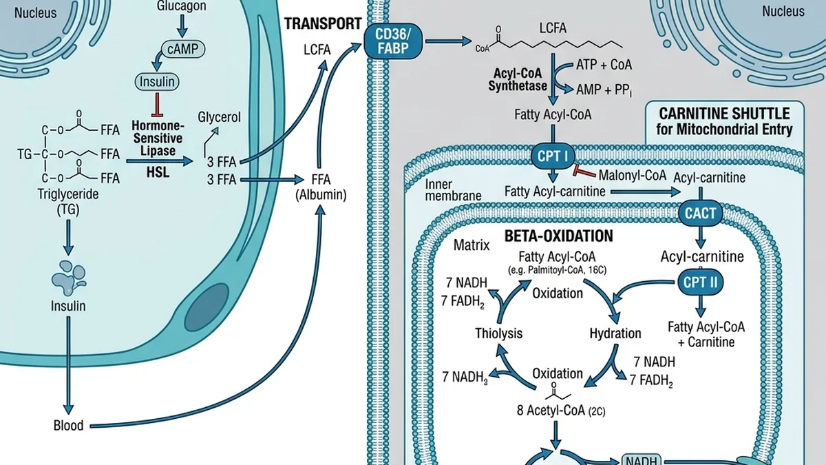
As you cross the 12-hour threshold, hepatic glycogen reserves become significantly depleted, falling by 50 to 80 percent. To maintain systemic energy supplies, the endocrine system initiates a major metabolic shift. The decrease in insulin relieves the inhibition on Hormone-Sensitive Lipase (HSL) in adipose tissue. HSL, activated by catecholamines (epinephrine and norepinephrine) binding to beta-adrenergic receptors, begins lipolysis: breaking down stored triglycerides in fat cells into free fatty acids (FFAs) and glycerol. These free fatty acids enter the Circulation and are transported to the liver and skeletal muscle tissues. Inside the hepatocytes, the enzyme Carnitine Palmitoyltransferase-1 (CPT-1) facilitates the transport of fatty acids across the mitochondrial membranes, initiating beta-oxidation. This process breaks down fatty acids into acetyl-CoA, which enters the Krebs cycle to produce ATP. When the liver is flooded with fatty acids, the production of acetyl-CoA exceeds the capacity of the Krebs cycle. The excess acetyl-CoA is directed toward ketogenesis (which can also be induced nutritionally using ratios from our free keto macros calculator), producing ketone bodies: acetoacetate and beta-hydroxybutyrate (BHB). Ketone bodies enter the bloodstream and cross the blood-brain barrier, providing a highly efficient alternative fuel source for the brain, preserving skeletal muscle mass by reducing the need for gluconeogenesis from amino acids.
Hours 18 to 24: The AMPK Trigger and Onset of Autophagy
This is the critical metabolic window where autophagy transitions from basal maintenance to active cellular repair. At approximately 18 continuous hours of zero caloric intake, hepatic glycogen is almost entirely exhausted. The cellular energy charge drops, resulting in a significant increase in the ratio of AMP to ATP. This alteration activates AMPK, which directly phosphorylates and shuts down the mTORC1 complex. Once mTORC1 is suppressed, the ULK1 complex is released, initiating the formation of the phagophore membrane. The cells, recognizing that no external nutrients are arriving, begin to seek out internal sources of energy and amino acids. They target dysfunctional components: misfolded proteins, oxidized lipid droplets, and damaged mitochondria that are leaking reactive oxygen species (ROS). The process of mitophagy (selective autophagy of mitochondria) is upregulated, allowing the cell to eliminate inefficient energy producers and replace them with healthy organelles. At 24 hours of fasting, systemic autophagy levels in tissues such as liver, muscle, and vascular endothelial cells show a significant increase, marking the onset of deep cellular repair.
Hours 24 to 48: Deep Autophagy and mTOR Shutdown
Between 24 and 48 hours of continuous fasting, your body enters a state of deep autophagy and metabolic adaptation. Circulating insulin levels reach their lowest physiological baseline, while glucagon, growth hormone, and cortisol are elevated to maintain blood glucose stability and preserve muscle tissue. In this state, the suppression of the mTOR pathway is complete, and the rate of autophagosome formation and lysosomal fusion reaches its peak. Recent clinical evidence from 2026 studies demonstrates that mitophagy and proteolysis (protein recycling) occur at high levels throughout the body, particularly in the liver, brain, and immune cells during this window. This deep recycling process clears out aggregated proteins associated with neurodegenerative decline, such as amyloid-beta and tau proteins. Additionally, the elimination of damaged mitochondria leads to a reduction in systemic oxidative stress and inflammation. The body is operating almost entirely on fat-derived ketones, with BHB levels rising to 1.5 to 3.0 mmol/L, providing neuroprotective signals and promoting the expression of Brain-Derived Neurotrophic Factor (BDNF) in the central nervous system.
Hours 48 and Beyond: Prolonged Fasting and Cellular Renewal
When a fast is extended beyond 48 hours, the cellular response transitions from recycling existing structures to initiating systemic regeneration. Clinical studies show that prolonged fasting (typically 48 to 72 hours) causes a significant reduction in circulating levels of Insulin-Like Growth Factor 1 (IGF-1) and downregulates Protein Kinase A (PKA) activity. This hormonal shift acts as a molecular trigger to clear out old, senescent immune cells (immunosenescence) through apoptosis. Once these worn-out white blood cells are removed, the reduction in IGF-1 and PKA signals hematopoietic stem cells in the bone marrow to transition from a dormant state to an active self-renewal program. Upon refeeding, these stem cells proliferate, generating fresh, highly functioning immune cells, effectively rejuvenating the immune system. However, extending fasts beyond 48 hours carries increased risks of electrolyte imbalances, muscle protein catabolism, and refeeding syndrome, and should only be conducted under close medical supervision.
2. What Happens Inside Your Cells: mTOR vs. AMPK
At the center of cellular lifespan regulation lies a delicate balance between two opposing proteins: Mechanistic Target of Rapamycin Complex 1 (mTORC1) and Adenosine Monophosphate-Activated Protein Kinase (AMPK). These two molecular complexes act as the fuel sensors of the human body, directing energy storage, cellular replication, protein synthesis, and lysosomal recycling. Under normal feeding conditions, the ingestion of macronutrients, particularly carbohydrates and amino acids, leads to a rapid elevation in circulating glucose and insulin. This insulin surge, combined with the presence of intracellular amino acids (specifically leucine, arginine, and lysine), activates the Rag GTPase complexes on the lysosomal membrane. This recruitment signals the activation of mTORC1, the master regulator of anabolic growth.
When mTORC1 is active, it promotes transcription of genes involved in ribosomal biogenesis, lipid synthesis, and cell growth. Concurrently, active mTORC1 directly phosphorylates and inactivates the ULK1 (Unc-51 Like Autophagy Activating Kinase 1) complex, specifically at the Serine 757 site. This phosphorylation event halts the assembly of the molecular machinery required to construct the autophagosome, the double-membrane vesicle that isolates cellular waste. As a result, in the fed state, autophagy is suppressed to a minimal basal level. The cell focuses entirely on expansion and synthesis, ignoring the accumulation of intracellular damage, oxidized proteins, and worn-out organelles. This state is metabolically optimal during periods of nutrient abundance, but when sustained chronically without relief, it leads to the accumulation of damaged mitochondria and senescent proteins, which accelerate aging and increase metabolic dysfunction.

Ganesh G Kamble
Ganesh G. Kamble is the founder and editor of Health is Heaven. He built this site after losing his father to a preventable condition, with the mission of providing clear, trackable health indicators so others can act early. His background is in the IT industry (16 years as an enterprise consultant) and he is not a medical professional; all medical content, formulas, and guides are reviewed by a credentialed medical reviewer/board before publication.